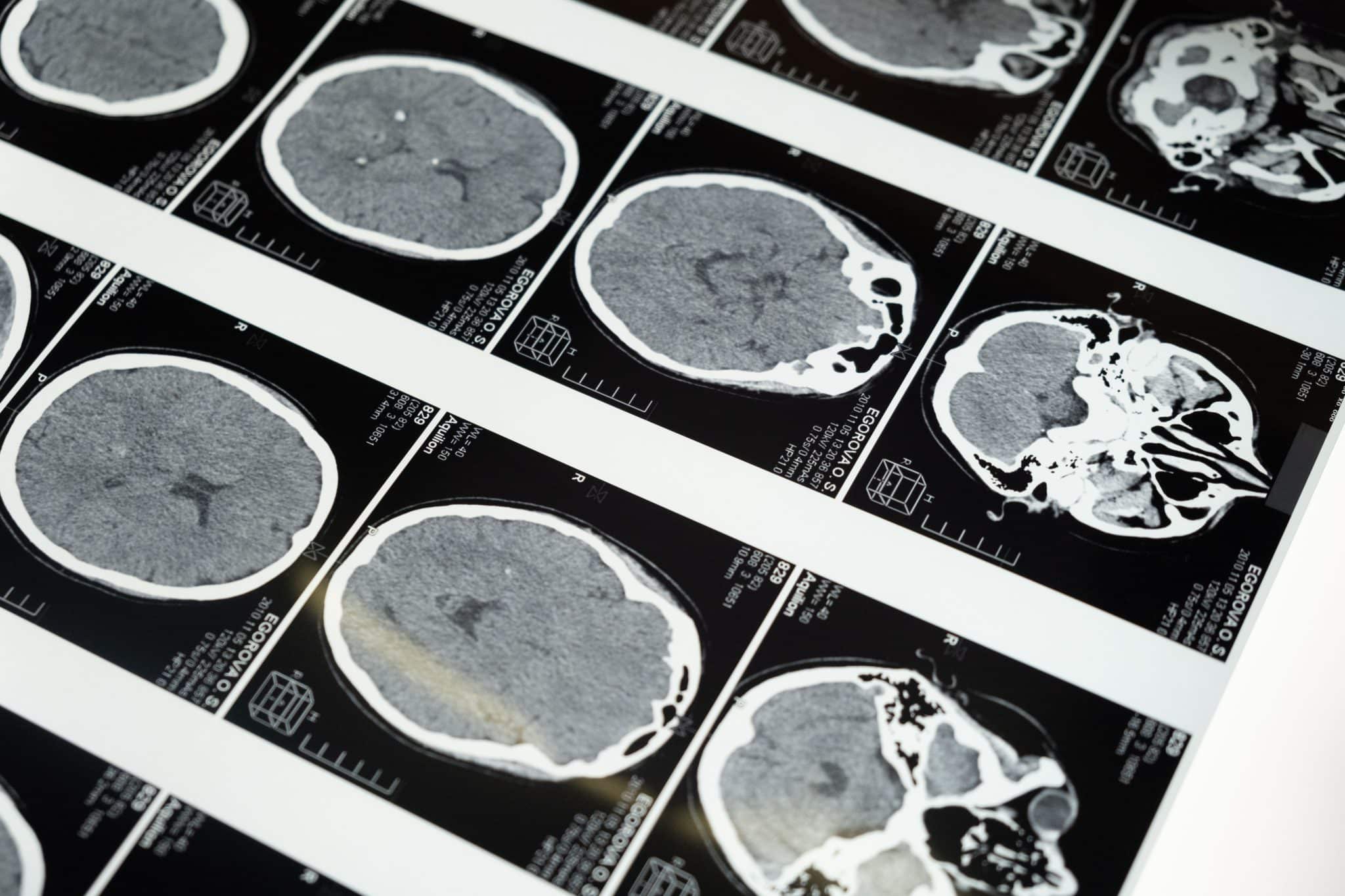

You hold your breath as the emergency room doctor walks in holding a tablet. After hours of waiting following your child’s terrifying fall, you finally hear the words every parent prays for: “The CT scan is clear.
You can take them home.” The immediate relief washes over you. You buckle your child into their car seat, trusting the medical technology that just gave you a clean bill of health.
But a few days or weeks later, a lingering sense of dread starts to creep in.
Your child just isn’t acting like themselves. They might be unusually fussy, struggling to balance, or sleeping far more than normal. Your parental intuition is screaming that something is wrong, yet you feel entirely dismissed by that initial “normal” medical report.
You are not alone in this terrifying gray area. Emergency departments treat approximately 700,000 children annually for traumatic brain injuries, mostly resulting from falls and sports.
Many of these families are sent home with a false sense of security.
The hard truth is that a normal emergency room CT scan does not guarantee safety. Early pediatric brain trauma often hides behind a deceptive symptom-free window, masking microscopic damage that standard hospital imaging simply cannot see.
Why a “Normal” CT Scan Doesn’t Guarantee Safety
To understand why your child might still be injured despite a clear scan, you need to understand how emergency rooms operate. The primary goal of emergency medicine is triage.
When a doctor orders a CT scan after a fall, they are urgently looking for immediate, life-threatening conditions.
They need to know if there is a massive brain bleed that requires emergency surgery or a skull fracture that threatens the brain’s structural integrity.
A CT scan is essentially a series of X-rays. It is fantastic at capturing dense material like bone and large pools of blood. However, it completely fails to capture the finer, microscopic details of the brain’s soft tissue network.
This limitation becomes dangerous when a child suffers a Diffuse Axonal Injury (DAI). A DAI occurs when the brain rapidly shifts inside the skull during a fall or sudden impact.
This violent sloshing motion shears and tears the brain’s microscopic nerve fibers, known as axons. Because these tears are microscopic, they are completely invisible on standard emergency room imaging.
The medical data on this diagnostic failure is staggering. Approximately 75% of patients with clinical signs of Diffuse Axonal Injury initially have normal CT scans but show abnormalities on an MRI.
This means three out of four patients are told their initial scans are clear while carrying a hidden, life-altering brain injury.
While an emergency room CT scan is great for ruling out immediate, life-threatening skull fractures, it is not designed to detect the microscopic nerve shearing that causes long-term cognitive deficits. If you suspect your child’s developing brain has suffered hidden trauma, consulting with a specialized brain injury attorney for children can help you secure the advanced neuroimaging and specialized medical evaluations your family needs before critical evidence is lost.
The “Silent Window” and the Danger of the Lucid Interval
One of the most confusing parts of a pediatric head injury is the immediate aftermath. Many children will cry intensely right after a fall, eventually calm down, and then return to playing with their toys.
This rapid return to “normal” behavior lulls parents and caregivers into believing the danger has passed.
In the medical community, this deceptive period is well-documented. For example, symptoms of an intracranial hematoma may develop right away or take weeks to appear, a period known as the “lucid interval.” During this silent window, the brain is actively experiencing internal swelling or slow bleeding, but the outward signs have not yet reached a tipping point.
Beyond the lucid interval, a young child’s brain has a unique biological way of hiding trauma. Infants and toddlers do not yet rely on higher-order logic or the executive function centers of the brain. The frontal lobe, which governs complex problem-solving, impulse control, and emotional regulation, is largely dormant during these early years.
Because the child is not yet performing complex tasks, the neurological deficits remain entirely hidden.
Think of it like a bridge under construction. If a support beam in the center of the bridge cracks, you will not know there is a problem until a heavy truck actually tries to drive over it. The injury is there, but the functional demand hasn’t exposed it yet.
Delayed Symptoms: What Parents Need to Watch For
Because of this silent window, parents must become vigilant observers in the weeks, months, and even years following a fall. Trust your gut. If your child seems “off,” do not let a previously clear CT scan stop you from seeking further medical advice.
Here are the early physical warning signs that may surface days or weeks later:
- Unexplained clumsiness or new issues with balance and coordination.
- Sudden, out-of-character irritability or inconsolable crying.
- Changes in sleep patterns, such as sleeping significantly more or struggling to stay asleep.
- A newly developed sensitivity to bright lights or loud noises.
- Changes in eating habits or frequent bouts of unexplained nausea.
As your child grows, the demands on their brain will increase. This is when you must look for long-term cognitive and behavioral signs:
- Missing expected developmental milestones at the 6, 12, or 18-month marks (e.g., delays in walking or speech).
- Sudden struggles with emotional regulation or severe behavioral outbursts in social settings.
- Academic regression, particularly around 3rd or 4th grade when school shifts from simple memorization to complex reading comprehension and independent problem-solving.
- Difficulty holding attention on a single task or following multi-step instructions.
The medical community recognizes this delayed onset as a major risk factor.
“Because of possible delayed presentation and high risk for problems with new learning, memory, attention, executive function, and language, TBI in this age group is viewed as a chronic disease process requiring ongoing monitoring and evaluation.”
How to Expose Hidden Trauma: Advanced Diagnostic Tools
If you are noticing any of the delayed symptoms listed above, your first step is to schedule an immediate follow-up with a pediatric neurologist. You must move past the emergency room doctors and consult with specialists who focus entirely on the brain’s long-term function.
When you speak to the neurologist, you have the right to request advanced neuroimaging alternatives. Since we know standard CT scans easily miss cellular damage, you need tools that look at the brain in entirely different ways.
The two most effective tools for exposing hidden brain trauma are the Magnetic Resonance Imaging (MRI) scan and Diffusion Tensor Imaging (DTI).
While an MRI uses powerful magnets to create highly detailed images of soft tissue and can spot subtle bruising, a DTI goes even further.
Diffusion Tensor Imaging is a highly advanced form of an MRI. It specifically maps the brain’s white matter tracts by tracking the movement of water molecules along the nerve fibers. In simple terms, if the microscopic nerves in the brain were sheared during a fall, the DTI will visually expose exactly where the water flow is disrupted.
It proves the existence of microscopic damage that a standard scan completely ignores.
| Diagnostic Tool | How It Works | What It Is Best For | Limitations |
|---|---|---|---|
| CT Scan | Uses X-rays to create cross-sectional images. | Ruling out immediate skull fractures and massive, life-threatening bleeds. | Cannot see microscopic nerve shearing or mild traumatic brain injuries. |
| Standard MRI | Uses strong magnetic fields to map soft tissue. | Detects subtle bruising, smaller bleeds, and inflammation. | May still miss the finest cellular damage in the brain’s white matter. |
| DTI (Diffusion Tensor Imaging) | Tracks water molecule movement along nerve pathways. | Exposes Diffuse Axonal Injury (DAI) and microscopic white matter damage. | Requires highly specialized equipment and an expert radiologist to interpret. |
The Hidden Clock: Legal Deadlines and Forensic Evidence
Watching your child for delayed symptoms is a medical necessity, but waiting for those obvious symptoms to appear before taking action carries severe legal and financial risks. If your child’s fall occurred at a daycare facility, a public school, or under a caregiver’s supervision, there is a hidden clock ticking against your family.
Forensic evidence rapidly decays or is routinely purged during your child’s symptom-free lag period. Surveillance footage from a daycare playground is often automatically overwritten every 30 days.
Incident reports can mysteriously disappear. Even broader medical records—like hospital admission logs or historical fetal heart monitoring strips in birth-related injury cases—can become difficult to track down as time passes.
If you wait a year to see if your child misses a developmental milestone, the physical proof of negligence may already be gone.
Furthermore, the legal system sets strict traps for families who wait too long. Depending on your state, “Statutes of Repose” can permanently bar you from filing a claim after a certain number of years, regardless of when you discovered the injury.
Even more urgently, if the fall happened at a public school or city-run facility, you may be subject to a “Notice of Claim” window. This law often requires you to formally notify the government entity of your intent to sue within a shocking 60 to 90 days of the accident.
Taking swift action early is the only way to secure the funding required to capitalize on pediatric neuroplasticity. A young brain has an incredible ability to adapt and rewire itself around damaged areas, but this window closes as the child ages. Early legal intervention preserves the evidence needed to win your case, allowing your family to pay for aggressive, specialized occupational and cognitive therapies while recovery is still highly possible.
Conclusion
The relief of a normal CT scan is entirely valid, but it should never be the final word on your child’s health. That initial emergency room visit simply ruled out the worst-case, immediate scenarios. It did not guarantee that your child’s developing brain was free of functional or microscopic trauma.
You know your child better than any doctor or medical machine. If your parental intuition is sounding the alarm, and you feel your child is acting differently in the days or weeks following a fall, you must pursue advanced medical evaluations immediately. Do not let a clear scan silence your protective instincts.
Protecting your child’s future requires a proactive approach both medically and legally. Seek out a free, no-risk consultation with a specialized legal team today. They can review your case, immediately secure vanishing evidence, and help connect you with top-tier pediatric neurologists.
Taking this step ensures your child gets the long-term financial and medical security they deserve, giving their brain the absolute best chance to heal.